(Available only at our Altrincham practice)
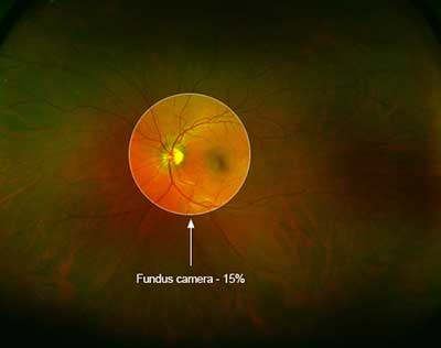
Every eye examination includes standard retinal imaging. The first image is a conventional photograph taken by a high-end digital camera that is focused on the retina at the back of the eye.
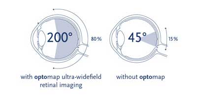
It typically provides your optometrist with a 45° view of the retina – though this can be more restricted with smaller pupils. The optic nerve and the macula are the main details that are seen in these photos. This allows us to detect and monitor any changes that may be indicative of glaucoma or macular degeneration.
The Optomap provides a much larger field of view of the retina, covering up to a 200° view of the retina.
This is generally not affected by pupil size so allows a thorough examination of the peripheral retina even with small pupils. In addition to allowing us to monitor and detect the same changes that can be seen with the conventional camera, Optomap’s wider field of view allows earlier detection of many systemic conditions such as high blood pressure, diabetes, high cholesterol levels, diabetic retinopathy and retinal tears/detachment.
For more information, click here to visit the Optomap website.


